“Post COVID-19 Lung Injury: What We Know So Far - Medscape” plus 3 more
“Post COVID-19 Lung Injury: What We Know So Far - Medscape” plus 3 more |
- Post COVID-19 Lung Injury: What We Know So Far - Medscape
- Biomarkers for acute respiratory distress syndrome | JIR - Dove Medical Press
- Vitamin D supplementation and Covid‐19 outcomes: A systematic review, meta‐analysis and meta‐regression - Wiley
- Acute Respiratory Distress Syndrome during the COVID-19 Pandemic - The Financial Express
| Post COVID-19 Lung Injury: What We Know So Far - Medscape Posted: 28 Jun 2021 07:48 AM PDT With vaccination rates increasing and new infections declining, we all hope the worst of the COVID-19 pandemic is over (fingers crossed really tight). Regardless, the post–COVID-19 syndrome pandemic has already begun. What is post–COVID-19 syndrome (or long-haulers or long-COVID)? Is it standard postviral fatigue? Prolonged deconditioning following debilitating illness? Permanent lung or vascular injury? Common sense and past experience say it's all of these. 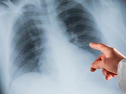 In theory, the burden of actual lung injury post-COVID-19 should be the easiest to quantify, so let's discuss what we think we know. I've heard experts break post–COVID-19 lung injury into three broad categories:
These categories are necessarily imprecise, making it challenging to fit some patients neatly into a single definition. For patients in the first category, management will largely be dictated by the nature of the pre-existing lung disease. For those in category two, we already know a lot about what their recovery from ARDS will look like. There's no longer reason to believe that COVID-19–related ARDS is particularly unique, and all things being equal, lung recovery should mimic that seen with non–COVID-19 ARDS. It's going to take patience and time, and beyond targeted rehabilitation it's not clear that we have anything available to expedite the process. The third category of patients is the most intriguing. Is there a group of patients who have residual lung injury but didn't have evident ARDS/ALI during their acute COVID-19 infection? Anecdotally we think so, but we know little about prevalence and less about management. A recent study published in Annals of the American Thoracic Society addresses both issues. In an observational report on patients recovering after being hospitalized with COVID-19 infection, the authors found that 3.6% of patients had residual lung injury that improved with 3 weeks of corticosteroid treatment. The report is timely and helpful but hardly definitive. It's observational, and patients required extensive screening and identification by a multidisciplinary committee of experts in interstitial lung disease. Patients were diagnosed as having organizing pneumonia (OP) as their "lung injury" if certain radiographic criteria were met. There were no biopsies. Last, there was no control group. Still, this report is critically important. It tells us that at 6 weeks post-discharge, about 3.6% of patients who were hospitalized for COVID-19 will have persistent symptoms, radiographic abnormalities, and a plateau in their recovery. Beyond that, it tells us little. Did these patients really have OP? It's impossible to know. The CT findings used to establish the diagnosis are nonspecific. Response to steroids is consistent with OP, but the treatment course was quite short. If truly OP, one would expect a high relapse rate after steroid withdrawal. Patients weren't followed long enough to monitor recurrence rates. Also, as appropriately discussed in the accompanying editorial, there's no control group so we can't know whether the patients treated with steroids would have recovered without treatment. There was objective improvement in lung function for the two to three patients they followed who did not receive steroids. However, it was of lesser magnitude than in the steroid group. Post–COVID-19 symptoms will remain a challenge for the foreseeable future. More than 30 million patients have been diagnosed with COVID-19 in the United States and close to half will experience persistent dyspnea. Putting the numbers together, I conclude that the vast majority will not have identifiable lung injury that will benefit from steroids. I wish I could prescribe patience to both physicians and patients. Aaron B. Holley, MD, is an associate professor of medicine at Uniformed Services University and program director of pulmonary and critical care medicine at Walter Reed National Military Medical Center. He covers a wide range of topics in pulmonary, critical care, and sleep medicine. Follow Medscape on Facebook, Twitter, Instagram, and YouTube | ||||||||||||||||||||||||||||||||||||||||||||||||||||||||||||||||||||||||||||||||||||||||||||||||||||||||||||||||||||||||||||||||||||||||||||||||||||||||||||||||||||||||||||||||||||||||||||||||||||||||||||||||||||||||||||||||||||||||||||||||||
| Biomarkers for acute respiratory distress syndrome | JIR - Dove Medical Press Posted: 01 Jun 2021 12:00 AM PDT IntroductionSepsis is a syndrome in which the body's immune system overreacts to infection. There may be several life-threatening complications. One of them is acute respiratory distress syndrome (ARDS).1,2 ARDS is an acute diffuse lung injury with high incidence. It has been shown that the pathophysiological basis of ARDS includes an excessive and protracted systemic inflammation,3 but the exact mechanisms remain unknown. The main risk factors that may cause ARDS include drinking, smoking, air pollution, hypoproteinemia, and diabetes mellitus.4–6 Currently, urgent treatment strategies mainly include low tidal mechanical ventilation, but the outcome is very poor.3 Although some progress has been made in treatment options in recent years, the mortality rate remains high.7 Previous studies have identified some genes used as biomarkers for ARDS. The high expression of interleukin (IL)-33 can elevate the expression of matrix metallopeptidases (MMP) 2 and 9 in patients with acute lung injury.8 SLC2A6 may be a critical biomarker for predicting survival of sepsis patients.9 V-set immunoglobulin-domain-containing 4 (VSIG4) may be involved in lung injury by inducing phagocytosis.10 Activation of ERK1/2 by NGF1B depends on MAPKKK c-Raf, eventually inducing inflammation. The NGF1B in the cytoplasm may be involved in the occurrence and development of ARDS.11 Down-regulation of the long non-coding RNA GAS5 decreases the expression of angiotensin-converting enzyme 2 (ACE 2), leading to increased levels of microRNA (miR) miR-200c-3p, which promotes the progression of ARDS.12 MYC and STAT3 may be the key regulatory genes in the underlying dysfunction of sepsis-induced ARDS.13 In addition, a previous study showed that gene set variation index can be used the potential diagnostic tool for sepsis.14 And gene set variation index may be considered as the biomarker of bacterial and fungal sepsis.15 However, we found that a majority of previous studies mostly focused on single genes or molecules for ARDS. There are only a few studies involving in a set of genes related to ARDS. Several studies confirmed that alveolar macrophage transcriptional programs are associated with ARDS.16–18 We hypothesized that these alveolar macrophage-related ARDS-specific transcriptional programs may be also reflected in blood cells. To investigate our hypothesis in the present study, we identified genes upregulated both in alveolar macrophages and blood cells and constructed a corresponding protein–protein interaction network associated with host response to ADRS. We found that eight genes formed a core gene set, and that the core gene set variation score may be used as a circulating biomarker for ARDS. Materials and MethodsData Collection and ProcessingThe data was downloaded from the Gene Expression Omnibus (GEO) database (https://www.ncbi.nlm.nih.gov/geo/): GSE116560,16 GSE89953,19 GSE7629320 and GSE32707.21 GSE116560 and GSE89953 were based on GPL6883. GSE116560 was obtained from alveolar macrophages of 68 ARDS samples, while GSE89953 was obtained from peripheral blood monocytes of 26 ARDS samples. The two datasets were combined and then batch effects were removed using the sva routine in R.22 From GSE76293, based on GPL570, the gene profiles of neutrophils from 12 ARDS samples and 12 healthy controls were extracted for subsequent analysis. From GSE32707, based on GPL10558, the gene profiles of whole blood cells were used to perform the subsequent analysis, from 18 ARDS samples and 34 healthy controls. The data were normalized using the "normalizeBetweenArrays" function of the limma routine23 in R. The probe set was converted into gene symbols, according to the annotation information of the three platforms. The workflow of the study is shown in Figure 1.
Principal Component Analysis (PCA) and Screening Differentially Expressed Genes (DEGs)The ggbiplot routine24 in R was applied to perform the PCA.25 In GPL6883 datasets (GSE116560 and GSE89953), the DEGs in alveolar macrophages were screened in comparison with peripheral blood monocytes. These DEGs may reflect the response of host monocyte-macrophages to ARDS.18 In GSE76293, DEGs in ARDS neutrophils were screened compared to healthy controls, since neutrophils are the most frequent nucleated cell in whole blood. In GSE32707, we screened the DEGs in whole blood between ARDS patients and healthy controls. A total of the DEGs were screened using the limma routine in R. The cut-off criteria are a false discovery rate (FDR)-adjusted P < 0.05 and |log2 (fold-change)| > 1. In the study, the common upregulated genes were defined as circulating ARDS alveolar macrophage-related genes (cARDSAMGs). Gene Ontology (GO) and Kyoto Encyclopedia of Genes and Genomes (KEGG) Pathway Enrichment AnalysisTo explore the potential biological functions of the cARDSAMGs, GO and KEGG pathway enrichment analysis were performed using the clusterProfiler routine in R.26 GO and KEGG networks were clustered using the plug-in ClueGO26 in Cytoscape software.27 Differences associated with an FDR-adjusted P < 0.05 was considered statistically significant. Analysis of Core GenesUsing STRING database28 (https://string-db.org/) and KEGG pathway enrichment analysis, a protein–protein interactions (PPIs) network was constructed for the cARDSAMGs. If genes were involved in ARDS-related pathways of interest, we defined them as core genes in the network. The GOsemsim routine inR29 was applied to analyze the semantic similarities among GO terms for the cARDSAMGs, applying the following formula: Pi = -lg (p) * |log2 (fold-change)|, weight (W) value = semantic similarities * mean value of Pi. Gene Set Variation Analysis (GSVA) and Receiver Operating Characteristic (ROC) Curve AnalysisThe GSVA routine inR30 was applied to calculate the core gene set variation analysis (CGSVA) score for individual samples to assess the concerted functional behavior of the core gene set. To further evaluate the potential diagnostic value of the CGSVA score for ARDS, an ROC curve analysis was performed using the pROC routine in R.31 ResultsMultiple Genes Identified as cARDSAMGsPCA showed that global gene expression patterns could distinguish ARDS from controls in GSE116560, GSE89953 and GSE76293, but not in GSE32707 (Figure S1). There were 8522 DEGs in GSE116560 and GSE89953, 6064 in GSE76293, and 7713 in GSE32707 (Figure 2A). A set of 60 DEGs was overexpressed in all the datasets, defined as cARDSAMGs (Figure 2B). Moreover, we obtained the p.value and rank of 60 cARDSAMGs in above three datasets and the ranking based on the p.value (Table S1). PCA showed that the expression patterns of the cARDSAMGs could not distinguish ARDS from healthy controls (Figure S2). However, we found that cARDSAMGs can better distinguish ARDS from control compared with the global gene expression patterns (Figures S1 and S2).
Biological Significance of cARDSAMGsGO enrichment analysis showed that changes in expression involved mainly genes related to metabolism- and inflammation-related biological processes (Figure 3A). KEGG result showed that DEGs were mainly involved in pathways related to ARDS, such as Notch the tumor necrosis factor (TNF) and the tumor necrosis factor (TNF) signaling pathways, as well as complement and coagulation cascades (Figure 3B). In addition, we performed the enrichment analysis using the 60 top ranked up-regulated genes in each of the datasets. As shown in Figure S3, we found that the biological processes (BPs) and KEGG pathways were different in each of datasets. But most of them were related to immunity, inflammation and phagocytosis. Furthermore, we randomly selected 60 genes in the three datasets and thereby calculated the enriched GO term. We found that the enriched GO terms mostly were not significantly related with inflammation, immunity and phagocytosis, which further evidenced that the gene set-cARDAMGs was not simply a randomly overlapping set (Figure S4). The ClueGo analysis indicated that DEGs were clustered mainly in biological processes, such as bone resorption, collagen catabolic process, regulation of cartilage development, positive regulation of ATP biosynthetic process, negative regulation of IL-2 production and positive regulation of animal organ morphogenesis (Figure 3C). No significant clustering of KEGG pathways was observed, based on a threshold of FDR-adjusted P < 0.05.
Core Genes in the PPIs Network of ARDSWe constructed the PPIs network of cARDSAMGs based on the STRING database. There were 25 nodes and 189 interaction pairs (Figure 4A). Previous studies showed that some of the biological processes and pathways were related to ARDS, including immunity, inflammation and phagocytosis.32,33 Therefore, we selected the following ARDS-related pathways as pathways of interest: Notch, TNF and IL-17 signaling pathways, as well as complement and coagulation cascades. Subsequently, we identified eight cARDSAMGs as core genes involved in these pathways. The core genes included PTCRA, JAG1, C1QB, ADAM17, C1QA, MMP9, VSIG4 and TNFAIP3. In the complement and coagulation cascades, antigen-antibody complexes act on C1QA and C1QB. They further act on VSIG4, thereby inducing phagocytosis.34,35 In the TNF signaling pathway, TNF acts on TNF receptor 1 (TNFR1), ultimately inducing tissue remodeling, autoimmune pathology, neutrophil recruitment and immunity to extracellular pathogens.36,37 In the Notch signaling pathway, ADAM17 and JAG1 act on the cell surface receptor Notch, which in turn affects PTCRA. In the TNF signaling pathway, TNF binds to TNFR1, which signals to surface receptors, intracellular signaling and remodeling of extracellular matrix.38,39 In the IL-17 signaling pathway, the IL-17 family signals activate downstream pathways that include NF-kappaB, MAPKs and C/EBPs, which further induces the expression of chemokines, cytokines and antimicrobial peptides40,41 (Figure 4B). The semantic similarities among the GO terms of the eight core genes are ranked in Figure 4C, and the W value of core genes, which may suggest the importance of molecules in this dysfunction, are displayed in Figure 4D.
CGSVA Score May Be Used as a Circulating Biomarker for ARDSAs shown in Figure S5, the C1QA, C1QB, MMP9, PTCRA and VSIG4 may be serve as circulating diagnostic markers for ARDS. Their areas under the ROC curves (AUCs) were higher than 0.7 in the three datasets. In addition, the CGSVA score was higher in the ARDS group than in the control group in all datasets (Figure 5A). The expression of the core genes is shown in Figure 5A. AUC for CGSVA score was higher than 0.7 in all three datasets, which suggests that the score may be useful as a diagnostic marker for ARDS (Figure 5B). The score in macrophages from the alveolar lavage fluid (GSE116560 and GSE89953) and whole blood (GSE76293 and GSE32707) was higher in ARDS individuals than in healthy controls (Figure 5C).
DiscussionIn this study, the differences in gene expression were explored between whole blood and alveolar lavage in ARDS samples and healthy controls. We selected four gene expression datasets from three platforms, where we found a common set of 60 DEGs upregulated in ARDS patients compared to healthy controls. Some among them have been previously informed to be related to ARDS. For example, immunohistochemical results showed that CD163 is overexpressed in ARDS patients.42 The highly expressed IL-33 can drive expression of MMP9 and MMP2 by activating STAT3. The process mainly happens in alveolar macrophages for acute lung injury patients induced by lipopolysaccharide.8 Mutations in the ALOX5 gene appear to be most likely cause of inter-individual differences in ARDS progression.43 Subsequently, we performed a function enrichment analysis for cARDSAMGs. The results showed that these cARDSAMGs were involved mainly in biological processes and pathways related to immunity, inflammation, phagocytosis and nucleic acid turnover. Some of these pathways have been related with ARDS in previous studies. Production of IL-1 and low concentrations of anti-inflammatory cytokines, such as IL-10 and IL-1 receptor antagonist, in bronchoalveolar lavage fluid of early ARDS patients have been closely related to poor prognosis.44 Numerous studies have linked inflammation and phagocytosis to ARDS.32,33,45,46 Moreover, we constructed a protein–protein interaction network to analyze the relationship among genes and pathways. We focused on TNF, IL-17 and Notch signaling pathways, as well as complement and coagulation cascades. In complement and coagulation cascades, complement activation may induce the occurrence of phagocytosis,47 which plays an essential role in ARDS.48 TNF acts as an inflammatory factor, up-regulating the expression of surface receptors, promoting intracellular signaling and remodeling of the extracellular matrix.49 Some biomarkers are closely related to ARDS, including surfactant-related proteins and cytokines (IL-1, −2, −6, −8, −10 and −15, and TNF-α), as well as neutrophil activation markers (MMP9, leukotriene B4 and ferritin).32 In the IL-17 signaling pathway, IL-17A and IL-17B act through surface receptors IL-17-17RA and IL-17RC to induce immunity and recruit neutrophils.49,50 Neutrophil-derived mediators induce epithelial cell death by oxidation of soluble Fas ligand.33,45 Eight genes corresponding to the selected pathways were identified as core genes: PTCRA, JAG1, C1QB, ADAM17, C1QA, MMP9, VSIG4 and TNFAIP3. VSIG4 is the main part of phagocytic system: it rapidly removes C3-modified particles.10 MMP9 is a secreted zinc metallopeptidase.51 MMP9 mediates protein degradation in the extracellular matrix of alveolar epithelial cells, mainly in intercellular junction proteins and proteins that anchor cells to the basement membrane.52 In addition, MMP9 is enriched mainly in macrophages and neutrophils,53,54 and involved in the occurrence and development of emphysema and asthma.55,56 TNFAIP3 was identified as a gene whose expression is rapidly induced by TNF. The protein encoded by this gene has been shown to inhibit nuclear factor (NF)-kappa B activation as well as TNF-mediated apoptosis.57 PTCRA encodes a single channel type 1 membrane protein in mature T cells. This protein forms a T-cell pre-receptor complex together with CD3 and T cell receptor beta chain (TCRB) complexes, thereby regulating the early development of T cells.58–60 C1QB may be associated with the immune response after injury.61 In addition, CGSVA score may be used as a biomarker for ARDS patients. Furthermore, the CGSVA scores associated with ARDS both in whole blood and alveolar macrophages were significantly higher in ARDS patients than in healthy controls. This may suggest that these alveolar macrophage-related ARDS-specific transcriptional programs are also reflected in blood cells. Our study presents several limitations. Firstly, our predictions are based on bioinformatic analyses, and therefore further experimental verification is needed. Secondly, most of alveolar macrophage genes have a dynamic pattern, but we only select genes that are significantly up-regulated in all time point without the consider of time changes. Thirdly, the whole transcriptome sequencing data of macrophages in whole blood and alveolar lavage fluid are difficult to find, currently. Whether these 8 cARDSAMGs may be used as diagnostic markers needs to verify in a larger independent clinical sample. Fourthly, it is shown that the gene set has a diagnostic performance in each of datasets in this study. Therefore, we believe that this gene set is specific for ARDS to some extent. But whether it is specific enough to ARDS, any inflammation or infection, still needs to be further clarified. Finally, we need to further explore the upstream regulators of core genes to reveal in more detail the imbalance network in ARDS. ConclusionsWe identified a core gene set (PTCRA, JAG1, C1QB, ADAM17, C1QA, MMP9, VSIG4 and TNFAIP3) that may help predict ARDS. The ARDS alveolar macrophage-related CGSVA score may be helpful for the diagnosis of ARDS. Data Sharing StatementThe data was downloaded from the Gene Expression Omnibus (GEO) database (https://www.ncbi.nlm.nih.gov/geo/): GSE116560, GSE89953, GSE76293 and GSE32707. FundingThis study was supported by the National Natural Science Foundation of China (81660132 and 81960343), the Reserve Cadre Training Program Science Foundation of the Second Affiliated Hospital of Guangxi Medical University (No. HBRC201805), Guangxi Health Commission key Laboratory of Emergency and Critical Medicine (The Second Affiliated Hospital of Guangxi Medical University) and the High-level Medical Expert Training Program of Guangxi "139" Plan Funding (G201903027). DisclosureThe authors declare that the research was conducted in the absence of any commercial or financial relationships that could be construed as a potential conflict of interest. References1. Zeng X, Feng J, Yang Y, et al. Screening of key genes of sepsis and septic shock using bioinformatics analysis. J Inflamm Res. 2021;14:829–841. doi:10.2147/JIR.S301663 2. Feng J, Pang J, He D, et al. Identification of genes with altered methylation and its role in early diagnosis of sepsis-induced acute respiratory distress syndrome. Int J Gen Med. 2021;14:243–253. doi:10.2147/IJGM.S287960 3. Peter JV, John P, Graham PL, Moran JL, George IA, Bersten A. Corticosteroids in the prevention and treatment of acute respiratory distress syndrome (ARDS) in adults: meta-analysis. BMJ. 2008;336(7651):1006–1009. doi:10.1136/bmj.39537.939039.BE 4. Matthay MA, Zemans RL, Zimmerman GA, et al. Acute respiratory distress syndrome. Nat Rev Dis Primers. 2019;5(1):18. doi:10.1038/s41572-019-0069-0 5. Ferguson ND, Frutos-Vivar F, Esteban A, et al. Clinical risk conditions for acute lung injury in the intensive care unit and hospital ward: a prospective observational study. Crit Care. 2007;11(5):R96. doi:10.1186/cc6113 6. Agrawal A, Matthay MA, Kangelaris KN, et al. Plasma angiopoietin-2 predicts the onset of acute lung injury in critically ill patients. Am J Respir Crit Care Med. 2013;187(7):736–742. doi:10.1164/rccm.201208-1460OC 7. Bellani G, Laffey JG, Pham T, et al. Epidemiology, patterns of care, and mortality for patients with acute respiratory distress syndrome in intensive care units in 50 countries. JAMA. 2016;315(8):788–800. doi:10.1001/jama.2016.0291 8. Liang Y, Yang N, Pan G, Jin B, Wang S, Ji W. Elevated IL-33 promotes expression of MMP2 and MMP9 via activating STAT3 in alveolar macrophages during LPS-induced acute lung injury. Cell Mol Biol Lett. 2018;23:52. doi:10.1186/s11658-018-0117-x 9. Li Z, Huang B, Yi W, et al. Identification of potential early diagnostic biomarkers of sepsis. J Inflamm Res. 2021;14:621–631. doi:10.2147/JIR.S298604 10. Helmy KY, Katschke KJ, Gorgani NN, et al. CRIg: a macrophage complement receptor required for phagocytosis of circulating pathogens. Cell. 2006;124(5):915–927. doi:10.1016/j.cell.2005.12.039 11. Jiang Y, Zeng Y, Huang X, et al. Nur77 attenuates endothelin-1 expression via downregulation of NF-kappaB and p38 MAPK in A549 cells and in an ARDS rat model. Am J Physiol Lung Cell Mol Physiol. 2016;311(6):L1023–L1035. doi:10.1152/ajplung.00043.2016 12. Li HB, Zi PP, Shi HJ, Gao M, Sun RQ. [Role of signaling pathway of long non-coding RNA growth arrest-specific transcript 5/microRNA-200c-3p/angiotensin converting enzyme 2 in the apoptosis of human lung epithelial cell A549 in acute respiratory distress syndrome]. Zhonghua Yi Xue Za Zhi. 2018;98(41):3354–3359. Chinese. doi:10.3760/cma.j.issn.0376-2491.2018.41.013 13. Zhang J, Luo Y, Wang X, et al. Global transcriptional regulation of STAT3- and MYC-mediated sepsis-induced ARDS. Ther Adv Respir Dis. 2019;13:1753466619879840. doi:10.1177/1753466619879840 14. Lu J, Li Q, Wu Z, et al. Two gene set variation indexes as potential diagnostic tool for sepsis. Am J Transl Res. 2020;12(6):2749–2759. 15. Zheng X, Luo Y, Li Q, et al. Two gene set variation index as biomarker of bacterial and fungal sepsis. Biomed Res Int. 2020;2020:8182358. doi:10.1155/2020/8182358 16. Morrell ED, Bhatraju PK, Mikacenic CR, et al. Alveolar macrophage transcriptional programs are associated with outcomes in acute respiratory distress syndrome. Am J Respir Crit Care Med. 2019;200(6):732–741. doi:10.1164/rccm.201807-1381OC 17. Thompson BT, Chambers RC, Liu KD, Drazen JM. Acute respiratory distress syndrome. N Engl J Med. 2017;377(6):562–572. doi:10.1056/NEJMra1608077 18. Aggarwal NR, King LS, D'Alessio FR. Diverse macrophage populations mediate acute lung inflammation and resolution. Am J Physiol Lung Cell Mol Physiol. 2014;306(8):L709–25. doi:10.1152/ajplung.00341.2013 19. Morrell ED, Radella F, Manicone AM, et al. Peripheral and alveolar cell transcriptional programs are distinct in acute respiratory distress syndrome. Am J Respir Crit Care Med. 2018;197(4):528–532. doi:10.1164/rccm.201703-0614LE 20. Juss JK, House D, Amour A, et al. Acute respiratory distress syndrome neutrophils have a distinct phenotype and are resistant to phosphoinositide 3-kinase inhibition. Am J Respir Crit Care Med. 2016;194(8):961–973. doi:10.1164/rccm.201509-1818OC 21. Dolinay T, Kim YS, Howrylak J, et al. Inflammasome-regulated cytokines are critical mediators of acute lung injury. Am J Respir Crit Care Med. 2012;185(11):1225–1234. doi:10.1164/rccm.201201-0003OC 22. Leek JT, Johnson WE, Parker HS, Jaffe AE, Storey JD. The sva package for removing batch effects and other unwanted variation in high-throughput experiments. Bioinformatics. 2012;28(6):882–883. doi:10.1093/bioinformatics/bts034 23. Cui Y, Yang X, Didelot X, et al. Epidemic clones, oceanic gene pools, and eco-LD in the free living marine pathogen vibrio parahaemolyticus. Mol Biol Evol. 2015;32(6):1396–1410. doi:10.1093/molbev/msv009 24. de Oliveira LA, da Silva CP, Nuvunga JJ, da Silva AQ, Balestre M. Bayesian GGE biplot models applied to maize multi-environments trials. Genet Mol Res. 2016;15(2). doi:10.4238/gmr.15028612 25. David CC, Jacobs DJ. Principal component analysis: a method for determining the essential dynamics of proteins. Methods Mol Biol. 2014;1084:193–226. doi:10.1007/978-1-62703-658-0_11 26. Yu G, Wang LG, Han Y, He QY. clusterProfiler: an R package for comparing biological themes among gene clusters. OMICS. 2012;16(5):284–287. doi:10.1089/omi.2011.0118 27. Kohl M, Wiese S, Warscheid B. Cytoscape: software for visualization and analysis of biological networks. Methods Mol Biol. 2011;696:291–303. doi:10.1007/978-1-60761-987-1_18 28. Szklarczyk D, Gable AL, Lyon D, et al. STRING v11: protein-protein association networks with increased coverage, supporting functional discovery in genome-wide experimental datasets. Nucleic Acids Res. 2019;47(D1):D607–D613. doi:10.1093/nar/gky1131 29. Yu G, Li F, Qin Y, Bo X, Wu Y, Wang S. GOSemSim: an R package for measuring semantic similarity among GO terms and gene products. Bioinformatics. 2010;26(7):976–978. doi:10.1093/bioinformatics/btq064 30. Hanzelmann S, Castelo R, Guinney J. GSVA: gene set variation analysis for microarray and RNA-seq data. BMC Bioinform. 2013;14:7. doi:10.1186/1471-2105-14-7 31. Robin X, Turck N, Hainard A, et al. pROC: an open-source package for R and S+ to analyze and compare ROC curves. BMC Bioinform. 2011;12:77. doi:10.1186/1471-2105-12-77 32. Sun X, Ma SF, Wade MS, et al. Functional promoter variants in sphingosine 1-phosphate receptor 3 associate with susceptibility to sepsis-associated acute respiratory distress syndrome. Am J Physiol Lung Cell Mol Physiol. 2013;305(7):L467–77. doi:10.1152/ajplung.00010.2013 33. Hogner K, Wolff T, Pleschka S, et al. Macrophage-expressed IFN-beta contributes to apoptotic alveolar epithelial cell injury in severe influenza virus pneumonia. PLoS Pathog. 2013;9(2):e1003188. doi:10.1371/journal.ppat.1003188 34. Bajic G, Degn SE, Thiel S, Andersen GR. Complement activation, regulation, and molecular basis for complement-related diseases. EMBO J. 2015;34(22):2735–2757. doi:10.15252/embj.201591881 35. Mathern DR, Heeger PS. Molecules great and small: the complement system. Clin J Am Soc Nephrol. 2015;10(9):1636–1650. doi:10.2215/CJN.06230614 36. Chu WM. Tumor necrosis factor. Cancer Lett. 2013;328(2):222–225. doi:10.1016/j.canlet.2012.10.014 37. Shuh M, Bohorquez H, Loss GE, Cohen AJ. Tumor necrosis factor-alpha: life and death of hepatocytes during liver ischemia/reperfusion injury. Ochsner J. 2013;13(1):119–130. 38. Hansson EM, Lendahl U, Chapman G. Notch signaling in development and disease. Semin Cancer Biol. 2004;14(5):320–328. doi:10.1016/j.semcancer.2004.04.011 39. Callahan R, Egan SE. Notch signaling in mammary development and oncogenesis. J Mammary Gland Biol Neoplasia. 2004;9(2):145–163. doi:10.1023/B:JOMG.0000037159.63644.81 40. Zenobia C, Hajishengallis G. Basic biology and role of interleukin-17 in immunity and inflammation. Periodontol 2000. 2015;69(1):142–159. doi:10.1111/prd.12083 41. Gaffen SL, Jain R, Garg AV, Cua DJ. The IL-23-IL-17 immune axis: from mechanisms to therapeutic testing. Nat Rev Immunol. 2014;14(9):585–600. doi:10.1038/nri3707 42. Maretta M, Toth S, Jonecova Z, et al. Immunohistochemical expression of MPO, CD163 and VEGF in inflammatory cells in acute respiratory distress syndrome: a case report. Int J Clin Exp Pathol. 2014;7(7):4539–4544. 43. Geiger EV, Doehring A, Kirchhof A, Lotsch J. Functional variants of the human 5-lipoxygenase gene and their genetic diagnosis. Prostaglandins Leukot Essent Fatty Acids. 2009;80(5–6):255–262. doi:10.1016/j.plefa.2009.04.001 44. Donnelly SC, Strieter RM, Reid PT, et al. The association between mortality rates and decreased concentrations of interleukin-10 and interleukin-1 receptor antagonist in the lung fluids of patients with the adult respiratory distress syndrome. Ann Intern Med. 1996;125(3):191–196. doi:10.7326/0003-4819-125-3-199608010-00005 45. Johnson ER, Matthay MA. Acute lung injury: epidemiology, pathogenesis, and treatment. J Aerosol Med Pulm Drug Deliv. 2010;23(4):243–252. doi:10.1089/jamp.2009.0775 46. Matthay MA, Ware LB, Zimmerman GA. The acute respiratory distress syndrome. J Clin Invest. 2012;122(8):2731–2740. doi:10.1172/JCI60331 47. Zilow G, Sturm JA, Rother U, Kirschfink M. Complement activation and the prognostic value of C3a in patients at risk of adult respiratory distress syndrome. Clin Exp Immunol. 1990;79(2):151–157. doi:10.1111/j.1365-2249.1990.tb05171.x 48. Hammerschmidt DE, Weaver LJ, Hudson LD, Craddock PR, Jacob HS. Association of complement activation and elevated plasma-C5a with adult respiratory distress syndrome. Pathophysiological relevance and possible prognostic value. Lancet. 1980;1(8175):947–949. doi:10.1016/s0140-6736(80)91403-8 49. Roca FJ, Whitworth LJ, Redmond S, Jones AA, Ramakrishnan L. TNF induces pathogenic programmed macrophage necrosis in tuberculosis through a mitochondrial-lysosomal-endoplasmic reticulum circuit. Cell. 2019;178(6):1344–1361 e11. doi:10.1016/j.cell.2019.08.004 50. Doyle MS, Collins ES, FitzGerald OM, Pennington SR. New insight into the functions of the interleukin-17 receptor adaptor protein Act1 in psoriatic arthritis. Arthritis Res Ther. 2012;14(5):226. doi:10.1186/ar4071 51. Nagase H, Barrett AJ, Woessner JF. Nomenclature and glossary of the matrix metalloproteinases. Matrix Suppl. 1992;1:421–424. 52. O'Kane CM, McKeown SW, Perkins GD, et al. Salbutamol up-regulates matrix metalloproteinase-9 in the alveolar space in the acute respiratory distress syndrome. Crit Care Med. 2009;37(7):2242–2249. doi:10.1097/CCM.0b013e3181a5506c 53. Kelly EA, Busse WW, Jarjour NN. Increased matrix metalloproteinase-9 in the airway after allergen challenge. Am J Respir Crit Care Med. 2000;162(3):1157–1161. doi:10.1164/ajrccm.162.3.9908016 54. Coussens LM, Tinkle CL, Hanahan D, Werb Z. MMP-9 supplied by bone marrow-derived cells contributes to skin carcinogenesis. Cell. 2000;103(3):481–490. doi:10.1016/s0092-8674(00)00139-2 55. Nakashima K, Hirota T, Obara K, et al. A functional polymorphism in MMP-9 is associated with childhood atopic asthma. Biochem Biophys Res Commun. 2006;344(1):300–307. doi:10.1016/j.bbrc.2006.03.102 56. Minematsu N, Nakamura H, Tateno H, Nakajima T, Yamaguchi K. Genetic polymorphism in matrix metalloproteinase-9 and pulmonary emphysema. Biochem Biophys Res Commun. 2001;289(1):116–119. doi:10.1006/bbrc.2001.5936 57. Das S, Pandey K, Kumar A, et al. TGF-beta1 re-programs TLR4 signaling in L. donovani infection: enhancement of SHP-1 and ubiquitin-editing enzyme A20. Immunol Cell Biol. 2012;90(6):640–654. doi:10.1038/icb.2011.80 58. Aifantis I, Borowski C, Gounari F, Lacorazza HD, Nikolich-Zugich J, von Boehmer H. A critical role for the cytoplasmic tail of pTalpha in T lymphocyte development. Nat Immunol. 2002;3(5):483–488. doi:10.1038/ni779 59. Del Porto P, Bruno L, Mattei MG, von Boehmer H, Saint-Ruf C. Cloning and comparative analysis of the human pre-T-cell receptor alpha-chain gene. Proc Natl Acad Sci U S A. 1995;92(26):12105–12109. doi:10.1073/pnas.92.26.12105 60. Saint-Ruf C, Lechner O, Feinberg J, von Boehmer H. Genomic structure of the human pre-T cell receptor alpha chain and expression of two mRNA isoforms. Eur J Immunol. 1998;28(11):3824–3831. doi:10.1002/(SICI)1521-4141(199811)28:11<3824::AID-IMMU3824>3.0.CO;2-9 61. Byrnes KR, Garay J, Di Giovanni S, et al. Expression of two temporally distinct microglia-related gene clusters after spinal cord injury. Glia. 2006;53(4):420–433. doi:10.1002/glia.20295 | ||||||||||||||||||||||||||||||||||||||||||||||||||||||||||||||||||||||||||||||||||||||||||||||||||||||||||||||||||||||||||||||||||||||||||||||||||||||||||||||||||||||||||||||||||||||||||||||||||||||||||||||||||||||||||||||||||||||||||||||||||
| Posted: 27 Jun 2021 12:00 AM PDT 1 INTRODUCTIONSevere acute respiratory syndrome coronavirus 2 (SARS-CoV-2) infection continues to spread globally causing a pandemic and has become a major medical focus for the last couple of years. The pandemic has involved over 164 million confirmed cases, with more than 3.4 million deaths as of 23 May 2021.1 While some SARS-CoV-2 infections appear as mild upper respiratory symptoms and may be self-limiting, notable numbers of patients require hospitalizations and intensive treatment following progression into a more severe cases, varying from simple lower respiratory tract infections to acute respiratory distress syndrome (ARDS) and eventually may turn into multi-organ failure.2, 3 Individuals with comorbidities such as obesity, diabetes, chronic respiratory disease, cardiovascular disease and other immunocompromising conditions are facing higher risks in developing the severe form of SARS-CoV-2 infections.4-9 For the past months, medical therapies to treat Covid-19 have been growing and evolving rapidly, ranging from supportive care, antivirals, anti-inflammatory agents and possible supplementations such as vitamin D.10-13 Several studies have shown that vitamin D has antiviral properties and potential roles against acute lung injury or ARDS, making large interest on vitamin D has rapidly emanated even in the early beginning of pandemic.14, 15 Low vitamin D serum levels is associated with an increase in inflammatory cytokine levels and significant increase of risk to develop pneumonia and viral respiratory tract infections, which both contribute to the development of ARDS.16-18 The goal of this study is to provide evidence whether or not vitamin D supplementation is associated with improved outcomes of Covid-19 based on the available studies. 2 MATERIALS AND METHODS2.1 Eligibility criteriaThe protocol of this systematic review and meta-analysis study of the observational and clinical trial studies was registered in PROSPERO (CRD42021256117). Included articles were selected as potentially fulfilling the entry criteria: comply the PICO framework (P: Covid-19 patients; I: vitamin D supplementation in any form; C: a group of patients who did not receive vitamin D, only receive standard of care therapy or any other medications as control/placebo; O: intensive care unit [ICU] admission, the need for mechanical ventilation and mortality), cohort, case-control, cross-sectional and randomized or non-randomized clinical trial articles were included. All studies other than original research articles (review articles, letter to editor or correspondence), case-series or case report studies, studies reported other than in English language, studies focussing on populations below 18 years of age and pregnant women were excluded. 2.2 Search strategy and study selectionThe papers from three databases (PubMed, Europe PMC and ClinicalTrials.gov) were searched systemically. Search terms used include 'vitamin D' OR 'calcidiol' OR 'calciferol' OR 'calcifediol' OR 'cholecalciferol' OR 'calcitriol' AND 'SARS-CoV-2', OR 'coronavirus disease 2019' OR 'Covid-19' in a time range from 2019 until 8 May 2021 with English-language restriction. Our searching strategy details are listed in Table 1. Initial screening of titles and abstracts was conducted to identify eligible articles. Searches of potential articles were also done by analysing the list of references of eligible studies. The search strategy was presented in the Preferred Reporting Items for Systematic Reviews and Meta-Analyses (PRISMA) diagram.
2.3 Data extraction and quality assessmentTwo authors (TIH and DI) performed the data extraction. An extraction form was developed to list the essential information about the study and its population characteristic (age, gender, hypertension, diabetes and corticosteroids usage/consumption), vitamin D dose, the number of patients receiving vitamin D and the control group, as well as the outcome of Covid-19 patients. The outcomes of interest are the rate of ICU admission, the need for mechanical ventilation and the mortality. The ICU admission rate is defined by the number of patients who were subsequently admitted into the ICU during the hospital stay. The need for mechanical ventilation is defined by the number of patients who need assisted ventilation. The total number of patients who were dead during the follow-up period with positive Covid-19 status was described as the mortality outcome. Two authors (JEH and HH) assessed the quality of each study included in this study independently. The quality of clinical trials was assessed using the modified Jadad scale assessment where the random allocation, allocation concealment, blindness and withdrawals and drop-outs of each study were evaluated. The studies were scored from zero to seven and a study ranked as a high-quality study if the score was >4.19 The quality of case-control and cohort studies were assessed using Newcastle–Ottawa Scale (NOS). The assessment reviews the selection, comparability and outcome of each study, then each study was assigned a total score from zero to nine. A study is graded as good quality if it scores ≥7. Cross-sectional studies was assessed using the Joanna Briggs Institute (JBI) Critical Appraisal Tools for Analytical Cross Sectional Studies. 2.4 Statistical analysisThe meta-analysis was performed using the Review Manager 5.4 (Cochrane Collaboration) software and Comprehensive Meta-Analysis version 3. Mantel–Haenszel's formula was employed to calculate odds ratio (OR) and its 95% confidence interval (95%CI) for the ICU admission outcome and the need for mechanical ventilation outcome, while Inverse Variance method was used to obtain the OR and 95% CI for the mortality outcome. The heterogeneity was assessed by using the I2 statistic with a value of <25%, 26–50% and >50% were considered as low, moderate and high degrees of heterogeneity, respectively. Random effects meta-regression was performed using a maximum likelihood for pre-specified variables including age, gender, hypertension, diabetes and the use of corticosteroids. The qualitative risk of publication bias was assessed with funnel plot analysis, while the quantitative risk of publication bias was assessed by using the Begg and Mazumdar rank correlation test.20 3 RESULTS3.1 Study selection and characteristicsThe searches on the databases yielded 6038 studies. A total of 5128 records remained following the elimination of duplicates. Screening the titles and abstracts and matching the inclusion and exclusion criteria, 5063 studies were removed. Among 65 evaluated full-text articles for its eligibility, 46 articles were excluded due to unavailable of results (still recruiting or withdrawn), 4 articles had no control or comparison group, 3 articles did not mention the criteria of the outcome of interest and 1 article because the full-text was not in English. The meta-analysis included 11 studies21-31 with a total of 2265 Covid-19 patients (Figure 1). Out of 11 studies, one study was double-blind randomized clinical trial, one study was open-label randomized clinical trial studies, four were retrospective cohort studies, two were prospective cohort studies and three were cross-sectional studies. All of the vitamin D doses were administered orally, although the dosage varied between each of the included studies, ranging from 25,000 IU/month up to 200,000 IU/day for two consecutive days. The baseline characteristics and severity between the vitamin D group and the control groups were already controlled in each of the included studies, meaning that there is no significant difference between two groups' characteristics. Table 2 presents the characteristics of the studies.  PRISMA diagram of the detailed process of selection of studies for inclusion in the systematic review and meta-analysis
3.2 Quality of study assessmentJadad scale assessments suggested that one clinical trial study was graded high quality, while another study was graded moderate quality (Table 3). Quality assessment of cohort and case-control studies using NOS scale and cross-sectional studies using JBI Critical Appraisal checklist indicated all included studies had a good quality (Table 4 and Table 5). Altogether, all studies were acceptable to be further analysed using meta-analysis.
3.3 Vitamin D and ICU admission of Covid-19 patientsFive studies (n = 772) reported the effect of vitamin D on the ICU admission of Covid-19 patients. Our pooled analysis showed that vitamin D supplementation was associated with reduction of ICU admission rate (OR 0.27; 95% CI: 0.09–0.76, p = 0.010, I2 = 70%, random-effect modelling; Figure 2a).  Forest plot that demonstrates the association of vitamin D supplementation with ICU admission rate (a), the need for mechanical ventilation (b) and mortality (c) outcomes 3.4 Vitamin D and the need for mechanical ventilationSix studies (n = 1516) reported the effect of vitamin D on the need for mechanical ventilation outcome. The pooled analysis suggested that vitamin D supplementation was associated with reduction of the need for mechanical ventilation (OR 0.34; 95% CI: 0.16–0.72, p = 0.005, I2 = 61%, random-effect modelling; Figure 2b). 3.5 Vitamin D and mortality of Covid-19 patientsTen studies (n = 2223) reported on mortality. The pooled estimate showed that vitamin D supplementation was associated with reduction of mortality from Covid-19 (OR 0.37; 95% CI: 0.21–0.66, p < 0.001, I2 = 50%, random-effect modelling; Figure 2c). 3.6 Meta regressionOur meta-regression suggested the association between vitamin D supplementation and mortality was affected by age (p = 0.027; Figure 3a), meaning that older people will gain more protective measures towards mortality in comparison with younger people. The association between vitamin D supplementation and mortality was not affected by gender (p = 0.191; Figure 3b), hypertension (p = 0.566; Figure 3c), diabetes (p = 0.608; Figure 3d), nor the use of corticosteroids (p = 0.070; Figure 3e).  Bubble-plot for meta-regression. Meta-regression analysis showed that the association between vitamin D supplementation and mortality outcome was affected by age (a), but not by gender (b), hypertension (c), diabetes (d) and the use of corticosteroids (e) 3.7 Publication biasFunnel plot analysis showed an asymmetrical inverted-plot for the ICU admission outcome (Figure 4a) and the need for mechanical ventilation outcome (Figure 4b), showing some indication of publication bias. Funnel plot analysis showed a relatively symmetrical inverted-plot for the mortality outcomes (Figure 4c). Begg and Mazumdar rank-correlation test were not statistically significant for ICU admission (p = 0.086), the need for mechanical ventilation (p = 0.060) and mortality outcome (p = 0.371), showing no indication of publication bias. However, because the number of included studies in the ICU admission and mechanical ventilation outcomes are fewer than 10 studies, the funnel plots and statistical tests for detecting publication bias are not very reliable when compared with larger numbers of included studies in each outcome.32, 33  Funnel plot analysis for the association of vitamin D supplementation with ICU admission (a), the need for mechanical ventilation (b) and mortality (c) outcomes 4 DISCUSSIONAccording to our pooled analysis, vitamin D supplementation had an association with a reduction of ICU admission rate, reduction in the need for ventilators and reduction of mortality from Covid-19. This meta-regression reveals that age affects the association between vitamin D supplementation and Covid-19 mortality. There are some explanations how vitamin D could affect the prognosis of Covid-19 patients. The interaction of SARS-CoV-2 infection and vitamin D are presented briefly in Figure 5. Both innate and adaptive arms of the immune system, triggered by SARS-CoV-2 infection, cause a destructive inflammatory reaction, resulting in local and systemic complications. The host immune or the inflammatory reaction is one of many severity-determining variables as proven by significant association between inflammatory proteins/indicators and disease severity.34, 35 Exhaustion markers (e.g., CD8+ T cells, NK cells, NKG2A) are increased during acute symptomatic period of Covid-19 and then return to normal in the convalescent period. Cytotoxic lymphocytes and natural killer cells aid control of viral infection, and both may serve as a predictor in determining the severity of the disease. Throughout the course of Covid-19 infection, Natural killer and cytotoxic lymphocytes will eventually reach functional exhaustion, as indicated by reduced total number.36 Severe Covid-19 patients have high levels of various inflammatory proteins such as C-reactive protein, D-dimer and cytokines, including IL-6, IL-1β, TNF-α, also known as cytokine storm.37 IL-6 can be used as a good indicator of poor outcome in Covid-19 patients who suffer ARDS.38, 39 Cytokine storm leads to a severe pulmonary infiltration by neutrophils and macrophages that causes severe alveolar injury with hyaline membrane formation and alveolar wall thickening.38 The cytokine storm increases inflammatory mediators and oxidative stress, while concomitantly reducing endothelial nitric oxide synthase. All these processes result in systemic inflammation and endothelial dysfunction, and ultimately cause hemodynamic instability, tissue injury and multiple organ failure.40  The role of vitamin D in suppressing the nuclear factor kappa B (NF-κB) signalling pathway (a) and the proposed potential therapeutic effect of vitamin D for COVID-19 and induced acute respiratory distress syndrome (b) There are three main mechanisms through which vitamin D may reduce the risk of infection: enhance physical barriers, cellular innate immunity and adaptive immunity. Vitamin D strengthens cellular immunity by induction of antimicrobial peptides, including human cathelicidin LL-37, and through 1,25-dihydroxyvitamin D and defensins.41 Cathelicidins are known to have direct antimicrobial effect towards a variety of pathogens comprising both Gram negative and positive bacteria, non-enveloped and enveloped viruses, and fungi.42 Cathelicidins also promote the chemotaxis of cellular immunity to the site of infection and its expression was upregulated by vitamin D especially in respiratory epithelial cells, which play a major role in host defenses.42 Furthermore, on the cellular level, vitamin D also stimulates gap junction genes, tight junction genes and adherens genes (e.g., E-cadherin) to strengthen cellular junction integrity and improve cell to cell communication, therefore maintaining the intercellular junctions to prevent further invasion of microorganisms, including viruses, which may lead to further inflammation and tissue damage.43, 44 The vitamin D receptor (VDR) is possessed by the majority of immune cells including macrophages, B and T lymphocytes, neutrophils and dendritic cells. VDR activation leads to downstream cell signalling that produces immunomodulatory, anti-proliferative, and pro differentiative effects.42 Vitamin D helps to reduce pro-inflammatory T helper 1 (Th1) cytokines, TNF-α and interferon (IFN)-γ and helps the production of Th2 lymphocyte cytokines which indirectly suppress Th1 cells.41, 45 Additional evidence that supports immunoregulatory function of vitamin D is that 1,25(OH)2D3 or calcitriol, the active form of vitamin D, is able to induce monocyte differentiation into macrophage-like form.42 VDR expressed by monocytes sensitizes them to the differentiating effects of calcitriol (autocrine mechanism for cell maturation). Vitamin D modulates macrophage response, and thus prevents overproduction of inflammatory cytokines and chemokines.42 Calcitriol downregulates granulocyte-macrophage colony stimulating factor but stimulates the immunosuppressive prostaglandin E2 production by the macrophages.41 Importantly, vitamin D deficiency impairs macrophage maturation.41 Several studies have shown that the risk of Covid-19 increases in people with vitamin D deficiencies; furthermore, lower concentration also contributes to the development of ARDS.46, 47 Aside from that, vitamin D has also been known to induce the production of type I IFNs, which able to suppress and keeping viral replication under control causing the prevention of further inflammatory response.48, 49 Several studies have reported that severe form of Covid-19 is frequently related with increased hypercoagulable state, which could be worsen by excessive inflammation and may accelerates upcoming thrombogenic events. Vitamin D has the potential role to promote antithrombin and thrombomodulin gene expression to suppress that hypercoagulable state, causing significant protection during the course of severe form of Covid-19.49-51 Vitamin D supplementation and serum levels above 50 ng/ml have been observed and may help in reducing severity and the course of viral diseases including Covid-19.41 Another role of vitamin D in the pathogenesis of Covid-19 is through its ability to inhibit the RAS and nuclear factor kappa B (NF-κB) pathway. Because ACE2 has a protective role against lung injuries, interference with that receptor by SARS-CoV-2 might be linked with ARDS. Lung injury caused by ischaemia and reperfusion are promoted by the increased expression of ACE/Ang II l and reduced levels of ACE2/Ang-(1-7). ACE2 plays its protective function to the lung through Ang-(1-7)/MasR pathway and prevents activation of NF-κB pathway/extracellular signal-regulated kinase.52 The 1α,25(OH)2D3, the biological active form of vitamin D, contributes its protective role and reduces lung injury by regulating RAS biosynthesis (renin, ACE/Ang II/AT1R axis and ACE2/Ang-(1-7) axis stimulation).53 The VDR and calcitriol also prevent lung, liver and kidney fibrosis through the downregulation of RAS and the inhibition of NF-κB and wnt/β-catenin.54, 55 A cohort study that investigates the influence of genetic variation in vitamin D pathway found that three SNP in VDR (rs4334089, rs11568820 and rs7970314) are associated with increased risk of upper respiratory tract infection.56 A systematic review and meta-analysis study which evaluates the association between VDR polymorphism and severe Respiratory Syncytial Virus (RSV)-bronchiolitis supports the association between FokI polymorphism and severe RSV infection. This study also examines the role of six VDR polymorphisms (Cdx, A1012G, FokI, BsmI, ApaI and TaqI) on infection susceptibility to enveloped virus found that a polymorphism at locus rs2228570 (FokI) is associated with viral infections.57 The TT genotype and T allele were reported to be risk factors for infections with enveloped viruses, including RSV.58, 59 Being an enveloped virus, it may be the same case for SARS-CoV-2. The mechanism that explains the association of FokI polymorphism with increased viral infection is that the FokI polymorphism creates a shorter VDR proteins that causes higher rate of transcription driven by the NF-κB, increased IL-12 and higher lymphocyte proliferation.56 Therefore, supplementation with vitamin D may help in ameliorating those potentially negative effects from viral infection. All of these properties and roles might explain the benefit of vitamin D on the outcomes of Covid-19. This study has some limitations. Significant heterogeneities were identified on most of the outcomes of interests included in this study. This was probably caused by the difference in the given vitamin D doses and co-administered medications with vitamin D as Covid-19 treatment. Importantly, we have made rigorous efforts to ensure that only sound studies were included, and several pre-print studies were included to minimize the risk of publication bias. 5 CONCLUSIONOur meta-analysis indicates that vitamin D supplementation had an association with favourable outcomes of Covid-19, compromising reduction in the rate of ICU admission, reduction in mechanical ventilation usage and reduction of mortality rate from Covid-19. This study suggests that vitamin D might be a potential therapeutic agent for the management of Covid-19 to give better outcomes for the patients. However, more randomized clinical trial studies are still necessary and should be done to confirming the results of our study. Finally, vitamin D might be considered as an essential drug for future Covid-19 therapy models. CONFLICT OF INTERESTThe authors declare that there is no conflict of interest. AUTHOR CONTRIBUTIONConceptualization, methodology, formal analysis, data curation, writing-original draft, visualization, writing-review and editing: Timotius Ivan Hariyanto. Conceptualization, methodology, formal analysis, data curation, writing-original draft, writing-review and editing: Denny Intan. Conceptualization, validation, supervision, writing-review and editing: Joshua Edward Hananto. Conceptualization, validation, supervision, writing-review and editing: Harapan Harapan. Conceptualization, validation, supervision, writing-review and editing: Andree Kurniawan. | ||||||||||||||||||||||||||||||||||||||||||||||||||||||||||||||||||||||||||||||||||||||||||||||||||||||||||||||||||||||||||||||||||||||||||||||||||||||||||||||||||||||||||||||||||||||||||||||||||||||||||||||||||||||||||||||||||||||||||||||||||
| Acute Respiratory Distress Syndrome during the COVID-19 Pandemic - The Financial Express Posted: 10 Jun 2021 12:00 AM PDT [unable to retrieve full-text content]Acute Respiratory Distress Syndrome during the COVID-19 Pandemic The Financial Express |


| You are subscribed to email updates from "respiratory distress syndrome,respiratory distress syndrome treatment,respiratory distress treatment" - Google News. To stop receiving these emails, you may unsubscribe now. | Email delivery powered by Google |
| Google, 1600 Amphitheatre Parkway, Mountain View, CA 94043, United States | |

Comments
Post a Comment